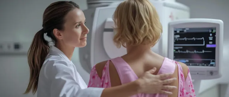
Breast Cancer Screening in Turkey - Tests and Recommendations
-
What Is Breast Cancer Screening?
-
Why Is Early Detection Important?
-
Breast Cancer Screening Recommendations
-
Types of Breast Cancer Screening Tests
-
Understanding Your Screening Results
-
Benefits and Risks of Breast Cancer Screening
-
Special Considerations in Breast Cancer Screening
-
Screening for Women with Dense Breasts
-
Addressing Disparities in Screening Outcomes
-
Talking to Your Doctor
-
Breast Cancer Screening at Liv Hospital
Regular breast cancer screening, like mammograms, aims to detect cancer early, often before symptoms appear, which significantly improves treatment success rates. It is a crucial tool for reducing mortality associated with this disease.
What Is Breast Cancer Screening?
The answer to the question 'What is breast cancer screening?' is quite detailed. Breast cancer screening is the process of checking a woman’s breasts for cancer before there are any signs or symptoms. The main goal of screening is to detect breast cancer at an early stage when it is more likely to be treated successfully. It’s important to understand that screening does not prevent cancer from developing; rather, it helps identify cancer early if it is already present. Early detection through screening can lead to better treatment outcomes and a higher chance of survival.

Why Is Early Detection Important?
Early detection breast cancer screening significantly improves treatment success rates and survival. When breast cancer is found at an early stage, it is often smaller and has not yet spread to other parts of the body, making it easier to treat. This allows for a wider range of treatment options, many of which may be less aggressive and have fewer side effects. Studies show that women diagnosed with early-stage breast cancer have much higher survival rates compared to those diagnosed at a later stage.
Annual breast cancer screening involves having a mammogram once every year and is often recommended for women at higher risk or those who prefer more frequent monitoring for earlier detection of potential abnormalities.
In short, early detection can save lives by allowing for timely and more effective treatment.
Breast Cancer Screening Recommendations
Breast cancer screening recommendations can vary slightly between different medical organizations, but there are some general consensus and key points to consider for women at average risk.
Breast cancer screening age recommendations provide guidelines on when women at average risk should begin and how often they should undergo screening mammography to detect the disease early.
Breast cancer screening guidelines provide recommendations for the use of various tests, primarily mammography, to detect breast cancer early in women at different risk levels and age groups.
The recommended breast cancer screening interval for most women is every 1 to 2 years, depending on age, risk factors, and medical guidelines.
Biennial breast cancer screening, which means screening every two years, is commonly recommended for women at average risk to balance the benefits of early detection with the potential risks of overdiagnosis breast cancer screening and false positives.
According to international recommendations such as those from the U.S. Preventive Services Task Force (USPSTF breast cancer screening) and the World Health Organization (WHO), women aged 50 to 74 are typically advised to have a mammogram every 1 to 2 years.
Some organizations, like the American Cancer Society, suggest offering screening as early as breast cancer screening age 40 based on individual risk and preference. Women with a higher risk—due to family history, genetic factors (like BRCA mutations), or prior chest radiation—may need to begin screening earlier and have additional tests like MRI.
They, cdc breast cancer screening and nci breast cancer screening are recommends mammography every 2 years for women aged 50-74 and suggests individualized discussions for women aged 40-49.
The breast cancer screening recommendation in Turkey suggests that women begin having regular mammograms starting at age 40, with screenings every 1–2 years depending on individual risk factors.
It's important that individuals consult with their healthcare provider to determine the most appropriate screening schedule for their personal risk profile.
The recommendations for when to start breast cancer screening vary slightly among different medical organizations, but here's a general overview for women at average risk:
Screening for Average-Risk Women
Women at average risk have no personal history of breast cancer, no known genetic mutation (like BRCA1/BRCA2), and no strong family history.
- Ages 40–49: Screening can begin based on individual choice, risk factors, and discussion with a healthcare provider.
- Ages 50–74: Mammogram every 1–2 years is strongly recommended by most international guidelines (e.g., USPSTF, WHO). Screening aims to detect cancer early, when it is more treatable and outcomes are better.
Screening for High-Risk Women
- High-risk women include those with: A strong family history of breast or ovarian cancer. Known BRCA1 or BRCA2 gene mutations. A personal history of radiation therapy to the chest (especially during youth)
- Screening often starts earlier (e.g., as early as age 30).
- Additional tests such as breast MRI may be recommended alongside mammography.
- A personalized screening plan should be developed in consultation with a specialist.
Screening After Age 75
Breast cancer screening older women typically continues until around age 75, depending on individual health and life expectancy. Routine screening after age 75 is not automatically recommended.
Decisions should be based on:
- Overall health status
- Life expectancy
- Personal values and preferences
For women in good health with a longer life expectancy, continuing screening may still be beneficial.

Types of Breast Cancer Screening Tests
Breast cancer screening tests to detect cancer early, often before symptoms appear. Mammograms, an X-ray of the breast, are the most common method. Clinical breast exams are done by a healthcare provider, while breast self-exams involve you checking your own breasts. Ultrasound uses sound waves to image the breast, helpful for evaluating lumps. MRI provides detailed images using magnets and is typically for high-risk individuals. Molecular breast imaging uses a tracer to highlight active cancer cells and can be useful for breast cancer screening dense breasts. A breast screening check-up is a routine examination to detect early signs of breast cancer, typically involving a mammogram and possibly a clinical breast exam.
Breast cancer screening methods include:
Mammography
Mammography breast cancer screening method that helps detect early signs of cancer, improving the chances of successful treatment. Mammography is the most widely used and recommended screening method for detecting breast cancer in women at average risk. It uses low-dose X-rays to capture images of the breast and can identify tumors or microcalcifications that may indicate early cancer, often before any symptoms appear. There are two types: 2D mammography, which captures flat images, and 3D mammography (digital breast tomosynthesis), which provides layered images for better accuracy, especially in women with dense breasts. Regular mammograms—typically every 1 to 2 years—have been shown to reduce breast cancer mortality by enabling earlier diagnosis and treatment.
Breast MRI
MRI breast cancer screening is a supplementary screening tool, often used for women at higher risk, to detect breast cancer with detailed imaging. Breast MRI (Magnetic Resonance Imaging) is a highly sensitive imaging method that uses magnetic fields and radio waves to create detailed pictures of the breast. It is primarily recommended for women at high risk of breast cancer, such as those with BRCA gene mutations or a strong family history.
MRI is usually used alongside, not instead of, mammography, as it can detect cancers that mammograms might miss. However, because of its sensitivity, it also has a higher rate of false positives, which may lead to unnecessary biopsies or anxiety.
Breast Ultrasound
Breast cancer screening ultrasound is often used as a supplemental tool alongside mammography, especially for women with dense breast tissue, as it can help detect abnormalities that may not be visible on a mammogram.
Breast ultrasound uses sound waves to produce real-time images of breast tissue and is often used as a supplementary screening tool. It is especially useful for evaluating specific areas of concern found on a mammogram or physical exam and for women with dense breast tissue, where mammograms may be less effective. Ultrasound can help distinguish between fluid-filled cysts and solid masses but is not typically recommended as a standalone screening test for the general population.
Clinical Breast Exam and Breast Self-Awareness
A clinical breast exam (CBE) is a physical examination of the breasts performed by a healthcare professional to check for lumps or other changes. While no longer universally recommended as a routine screening tool, it may still play a role in settings with limited access to imaging. Breast self-awareness encourages individuals to become familiar with the normal look and feel of their breasts, so they can notice any unusual changes early. Though not proven to reduce breast cancer mortality, self-awareness can empower women to seek timely medical evaluation.
Other Investigated Methods
Supplemental breast cancer screening refers to additional imaging tests used in conjunction with mammography for women at higher risk of breast cancer or those with dense breast tissue.
Several experimental or less commonly used methods are being studied for breast cancer screening. Thermography, which detects heat patterns on the breast surface, lacks sufficient evidence of accuracy and is not recommended by major health organizations. Molecular Breast Imaging (MBI) shows promise in detecting cancer in dense breasts but is not yet standard practice.
Blood-based tests and liquid biopsies are also under investigation as future tools for early cancer detection, though they are not currently approved for routine screening use.
Understanding Your Screening Results
After a breast cancer screening, your results can be either normal or abnormal. A normal result means no signs of cancer were detected, while an abnormal result indicates that further tests, such as an ultrasound or biopsy, may be needed to check for potential issues.
It's important to note that an abnormal result doesn’t necessarily mean cancer; it may be a false positive breast cancer screening, where the test suggests a problem that isn’t there, or a false negative breast cancer screening, where cancer is present but not detected. Always follow up with your healthcare provider to discuss results and any necessary next steps.
Breast cancer screening follow-up refers to the next steps taken after an initial screening, particularly if an abnormal result is found.
Normal Results
- No signs of cancer detected during the screening.
- Indicates healthy breast tissue at the time of the exam.
- You will likely continue with regular screenings (every 1–2 years), depending on your age and risk factors.
Abnormal Results and Follow-Up
Abnormal breast cancer screening results do not necessarily mean cancer is present but indicate that further testing is needed to evaluate any irregularities.
The next steps may include:
- Additional imaging tests (e.g., ultrasound or MRI) to look more closely at the area of concern.
- If these tests are still inconclusive or show signs of possible cancer, a biopsy may be recommended.
Biopsy involves removing a small sample of tissue to be examined under a microscope to check for cancer cells.
Most abnormal findings are not cancer, and the follow-up process helps ensure accurate diagnosis and timely treatment.

Benefits and Risks of Breast Cancer Screening
Breast cancer screening offers important benefits, including the early detection of cancer, which can lead to higher survival rates, less aggressive treatment, and more options for care. However, screening also carries some risks, such as false positives that can lead to anxiety and unnecessary tests, false negatives that may delay diagnosis, and the possibility of overdiagnosis - detecting cancers that might never have caused harm.
Additionally, mammograms involve low levels of radiation, though the risk is minimal. Understanding both the potential breast cancer screening benefits and risks helps individuals make informed decisions in consultation with their healthcare providers.
Benefits of Screening
Breast cancer screening helps detect cancer at an early stage, often before any symptoms appear, increasing the chances of successful treatment and survival. Early detection can lead to less aggressive treatments, more treatment options, and better overall outcomes. For many women, receiving a normal result also provides peace of mind and encourages continued attention to breast health.
Risks of Screening
While screening has clear benefits, it also involves some risks. False positives can cause unnecessary anxiety and lead to additional tests or biopsies that turn out to be harmless. False negatives may delay diagnosis by missing cancers that are actually present. Overdiagnosis is another concern, as some detected cancers may never cause harm but still result in treatment. Lastly, mammography involves exposure to a small amount of radiation, though the risk is considered very low.
Special Considerations in Breast Cancer Screening
Breast cancer screening is not one-size-fits-all—certain individuals may require tailored approaches based on their age, personal or family history, genetic factors, and breast density. Women with dense breast tissue may benefit from additional imaging, such as ultrasound or MRI, since dense tissue can make cancers harder to detect on mammograms.
Those with genetic mutations like BRCA1 or BRCA2, or a strong family history of breast or ovarian cancer, are considered high-risk and may need to begin screening earlier and undergo more frequent or advanced imaging. Cultural, social, and access-related factors can also affect screening participation and should be addressed to ensure equitable care.
Screening for Women with Dense Breasts
Breast density refers to the proportion of fibrous and glandular tissue compared to fatty tissue in the breast, as seen on a mammogram. Women with dense breasts have more glandular and connective tissue, which appears white on a mammogram—just like tumors—making it harder to detect cancer through standard mammography. Dense breast tissue is also associated with a slightly increased risk of developing breast cancer.
Because of these challenges, women with dense breasts may benefit from supplemental screening, such as ultrasound or breast MRI, to improve detection rates. It’s important for women to discuss their breast density with their healthcare provider to determine the most effective screening strategy.
Addressing Disparities in Screening Outcomes
Not all individuals have equal access to or outcomes from breast cancer screening. Disparities can arise due to differences in race, ethnicity, income, geographic location, education, and access to healthcare.
- Access to Care: Women in rural or underserved areas may face barriers such as fewer healthcare facilities, lack of insurance, or transportation challenges that limit access to screening.
- Socioeconomic Inequities: Lower income and education levels are often linked to reduced screening rates and delayed diagnoses.
- Racial and Ethnic Disparities: Black and Hispanic women are more likely to be diagnosed at later stages and may have poorer outcomes, often due to systemic inequities in healthcare access and quality.
- Cultural and Language Barriers: Limited health literacy, language differences, or cultural beliefs about illness can affect understanding and participation in screening programs.
- Solutions Needed: Addressing these disparities requires targeted outreach, improved access to affordable screening services, culturally sensitive education, and inclusive healthcare policies.
Talking to Your Doctor
Having an open conversation with your doctor is essential to making informed decisions about breast cancer screening. Your provider can help assess your individual risk based on personal and family history, lifestyle factors, and breast density. Together, you can determine when to begin screening, how often it should occur, and whether additional tests are needed. Don’t hesitate to ask questions or express concerns—shared decision-making ensures that your screening plan fits your unique needs and values.
Breast Cancer Screening at Liv Hospital
Liv Hospital offers a comprehensive and personalized approach to breast cancer screening, utilizing advanced diagnostic technologies and a multidisciplinary team of specialists. You can prevent your breast cancer risk with early diagnosis by having a health check up, genetic check up in the check up center section of our hospital.
- Advanced Imaging Techniques: Liv Hospital employs state-of-the-art imaging methods, including digital mammography with 3D tomosynthesis, breast ultrasonography, and magnetic resonance imaging (MRI), to detect breast abnormalities at the earliest stages.
- Specialized Care for Dense Breast Tissue: For women with dense breast tissue, which can make cancer detection more challenging, Liv Hospital offers supplemental screening options like automated breast ultrasound to enhance diagnostic accuracy.
- Genetic Risk Assessment: Individuals with a family history of breast or ovarian cancer can undergo genetic screening for mutations such as BRCA1 and BRCA2. This allows for personalized risk assessment and tailored screening strategies.
- Multidisciplinary Evaluation: Patients receive coordinated care from a team that includes general surgery specialists, radiologists, pathologists, oncologists, and genetic counselors. This collaborative approach ensures comprehensive evaluation and management of breast health .
- Biopsy Services: When imaging results indicate the need for further investigation, Liv Hospital provides minimally invasive breast biopsy procedures to obtain tissue samples for pathological examination.
- Customized Follow-Up Plans: Based on screening outcomes, Liv Hospital develops individualized follow-up programs, which may include regular monitoring or treatment planning, to ensure optimal patient care.
Our Approach to Screening and Diagnosis
At Liv Hospital, we take a patient-centered, evidence-based approach to breast cancer screening and diagnosis. Our goal is to detect breast abnormalities at the earliest and most treatable stage, using advanced technologies and personalized risk assessment. Each patient receives comprehensive care from a multidisciplinary team of specialists who guide them through every step—from screening to diagnosis and beyond.
Available Screening Technologies
We offer a full range of advanced imaging options to ensure accurate detection, including:
- 3D Digital Mammography (Tomosynthesis): Enhances image clarity and helps detect smaller tumors, especially in dense breasts.
- Breast Ultrasound (4D Breast Ultrasound): Used as a supplemental tool, particularly effective in evaluating dense breast tissue.
- Breast MRI: Recommended for high-risk individuals or when more detailed imaging is needed.
- Automated Breast Ultrasound (ABUS): Offers consistent, high-resolution images and supports early detection in women with dense breasts.
Schedule Your Breast Cancer Screening
Taking the first step toward breast health is easy at Liv Hospital. You can schedule your screening:
- Online through our official website
- By phone via our patient support line
- Or by visiting any Liv Hospital branch to speak directly with our care team
Our specialists are here to help you determine the best screening plan based on your age, risk level, and personal health history
Frequently Asked Questions
For most women at average risk, breast cancer screening with mammography is recommended to begin between ages 40 and 50, depending on personal and family health history. It’s best to consult your doctor to decide the right time for you.
Most guidelines recommend a mammogram every 1 to 2 years for women aged 50 to 74. Women in their 40s or those at higher risk may benefit from starting earlier and screening more frequently.
Mammograms can cause temporary discomfort or pressure as the breast is compressed during the imaging, but it’s usually brief. Pain levels vary by individual, and the process only takes a few minutes.
Dense breasts have more glandular and connective tissue than fatty tissue. This is common and not abnormal, but it can make it harder to detect cancer on a mammogram and is linked to a slightly higher risk of breast cancer.
An abnormal result doesn’t mean you have cancer. It means something unusual was seen, and more tests—like an ultrasound, MRI, or biopsy—may be needed to learn more. Most follow-up results are not cancerous, but further evaluation is important for your safety.
* Contents of this page is for informational purposes only. Please consult your doctor for diagnosis and treatment. The content of this page does not include information on medicinal health care at Liv Hospital


