


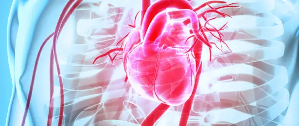
Cardiac MRI
The heart is one of the most difficult areas to image because it is located inside the rib cage and behind the bony structures and is in constant motion. Therefore, centers with advanced technology and experienced experts are required to examine their functions and diseases in detail. Cardiac MRI is an advanced imaging method that allows the heart to be examined in the most detail without the need for invasive procedures.
Cardiac MRI at Liv Hospital
Cardiac MRI (Magnetic Resonance) service provided by Liv Hospital is a modern medical service that offers detailed imaging and diagnostic methods related to the cardiovascular system. This service is used to evaluate the structural and functional characteristics of the heart and is an important tool for the diagnosis, planning and follow-up of heart diseases.
Cardiac MRI service at Liv Hospital is performed by expert cardiologists and experienced healthcare personnel. In this way, patients receive a detailed evaluation of their heart health and are also subjected to a reliable diagnosis and treatment process.
Can also be applied to children
Since no radiation is used in cardiac MR imaging, it can be performed safely in both children and adults. Again, the medication (contrast material) to be administered if necessary during the procedure is much safer than tomographic methods. With this examination method, the functions of the heart are calculated in the most accurate way. Heart structures can be examined in any plane, without angle limits. Pathologies in the heart are clearly revealed.
In Which Situations Is It Done?
It provides detailed information in detecting damage to the heart muscle in patients with occlusion in the heart vessels (Viability assessment). The rate of this damage is very valuable as it determines whether the patient will benefit from heart surgery.
It contributes greatly to preoperative planning and risk classification by allowing detailed evaluation of the heart and great vessels in congenital heart diseases. It also allows obtaining the most accurate and reliable data for lifelong follow-up after surgery.
It is used to determine the degree of disease in cases of stenosis or insufficiency of the heart valves. In these diseases, the rate of regurgitation of blood through the valves can be calculated with great accuracy.
It allows the underlying cause of heart rhythm disorders to be revealed.
It can be used to differentiate between mass and clot in patients with suspected heart mass, and to identify malignant tumors if there is a mass.
It allows the heart muscle to be evaluated in detail in diseases such as Mediterranean anemia (thalassemia), sarcoidosis, Anderson-Fabry disease, and amylodiosis, and to monitor the rate of damage to the heart.
In what cases is it not done?
It is not appropriate to perform an MRI examination on people who have a pacemaker or medical equipment (prosthesis, cochlear implant, etc.) that is not compatible with MRI. It should also be done without proper medical support for people with claustrophobia.
What kind of examination is heart MRI?
Before starting the examination, a vascular access is established in the arm. After the patient is taken to the MRI machine, ECG tapes are placed on the chest to monitor the movements of the heart. Since the patient needs to hold his breath from time to time during the examination, breath-holding practice is performed and MRI imaging is started. If necessary, a medicine called contrast material is administered through the vascular access. The review is completed on average between 30 and 45 minutes.
* Contents of this page is for informational purposes only. Please consult your doctor for diagnosis and treatment. The content of this page does not include information on medicinal health care at Liv Hospital

