


Detailed Ultrasound: What is it? When is it done?


-
Detailed Ultrasound: What is it?
-
When is Detailed Ultrasound Done?
-
The Importance and Advantages of Detailed Ultrasound
-
The Detailed Ultrasound Process
-
How Detailed Ultrasound Results are Evaluated
-
Detailed Ultrasound and Risk Assessment

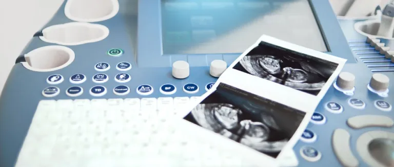
Detailed ultrasound is an imaging method that uses high-frequency sound waves to examine specific organs or systems in the body. This technology provides detailed images of organ structures and provides diagnostic information. It is commonly used for various medical purposes such as monitoring pregnancy, evaluating abdominal or pelvic organs, etc. Detailed ultrasound is a medical imaging method used to more thoroughly assess the development and health of the baby during pregnancy. Regarding the question of when detailed ultrasound is done, it is typically performed between the 18th and 22nd weeks of pregnancy and is often referred to as the "20-week ultrasound" or "pregnancy ultrasound.
Detailed Ultrasound: What is it?

Detailed ultrasound is an essential part of standard pregnancy monitoring. During this examination, the baby's body structure, organs, bones, gender, and other important anatomical details are examined using high-frequency sound waves. Compared to standard ultrasounds, detailed ultrasound provides a more thorough examination, allowing for the early detection of potential issues and the development of appropriate intervention plans. During this examination, the baby's heartbeat, brain development, kidneys, liver, and other organ systems are observed in detail. Additionally, whether the baby is at risk for congenital anomalies or genetic conditions is also evaluated.
When is Detailed Ultrasound Done?
Detailed ultrasound is typically performed between the 18th and 22nd weeks of pregnancy to more thoroughly assess the baby's health. During this examination, the baby's anatomical structures are examined in detail, potential issues can be detected early, and appropriate intervention plans can be made. This is important to support the baby's healthy development process. If you're wondering when the last week for detailed ultrasound is, it can be performed beyond the 22nd week if deemed necessary by the healthcare professional.
The Importance and Advantages of Detailed Ultrasound
Detailed ultrasound plays a crucial role during pregnancy as it enables healthcare professionals to detect potential issues with the baby's health early and initiate treatment. The main advantages of detailed ultrasound are as follows:
- Detailed ultrasound is used to thoroughly evaluate the baby's anatomical structure. Examining organs, bones, brain development, and other important anatomical details allows for the early detection of potential issues.
- This examination enables the early detection of congenital anomalies or developmental problems. Early diagnosis provides healthcare professionals with the opportunity to create appropriate treatment and management plans, which is essential for ensuring the baby's health.
- Detailed ultrasound is used to assess the potential effects on the baby's health in pregnancies with genetic or other risk factors. This informs the expectant mother and the healthcare team about potential risks.
- Expectant mothers often have the opportunity to learn the baby's gender during detailed ultrasound. This can contribute to emotional bonding and the preparation process for the family.
- Detailed ultrasound is an important tool for monitoring the health of both the mother and the baby during pregnancy. This examination is used to identify potential issues during pregnancy and determine appropriate treatment plans.

The Detailed Ultrasound Process
The process of detailed ultrasound is generally fast and painless. This examination allows for obtaining important information about the baby's health, enabling closer monitoring of the pregnancy process. The detailed ultrasound process consists of 5 stages:
- Preparation Stage: The expectant mother typically does not wear any special clothing, but the abdominal area is cleaned with gel to be used during the examination. This gel ensures better contact of the ultrasound probe with the skin.
- Application Stage: The expectant mother lies on her back, and a special gel is applied to the abdominal area by the ultrasound technician or doctor. Then, the ultrasound probe is maneuvered over the abdomen to ensure that the sound waves reach the organs.
- Imaging Stage: Images created by the reflections of sound waves are instantly displayed on a computer screen. During this stage, the baby's organs, bone structure, gender, and other important anatomical details are examined.
- Measurements and Evaluation Stage: Important parameters such as the baby's size, heart rate, and organ development are measured and evaluated. This stage is critical for determining whether the baby is developing healthily.
- Explanation of Results: After the detailed ultrasound, the obtained images and measurements are evaluated. The expectant mother is informed about the baby's health condition and development.
Throughout the entire process, if the healthcare professional observes any abnormalities, they may resort to additional tests and treatment methods.

How Detailed Ultrasound Results are Evaluated
Detailed ultrasound results play a critical role in the prenatal screening process. They contain important information about the baby's development. During the anatomical assessment, the baby's organs, bone structure, and gender are examined in detail. If any abnormalities or developmental issues are detected, they are communicated to the parents.
Biometric measurements are taken to assess the baby's growth rate and monitor overall development. Additionally, the baby's heart rate and circulatory system are examined to obtain important information about cardiovascular health. During this process, the condition of the expectant mother's uterus is also evaluated, ensuring whether the baby is developing in a healthy environment. Detailed ultrasound results enable expectant mothers to closely monitor the pregnancy process, facilitating the planning of necessary precautions and interventions.
Detailed Ultrasound and Risk Assessment
In detailed ultrasound, risk assessment is conducted considering factors such as the expectant mother's genetic background, age, and pregnancy history, along with the obtained data. Based on this assessment, potential risks are identified, and additional tests and treatments may be applied if necessary.
Frequently Asked Questions
Detailed ultrasound is considered a safe medical imaging method by experts and carries a very low risk during the pregnancy process.
If you're wondering when the results of a detailed ultrasound are obtained, they are typically available immediately after the examination, on the same day, or within a few days.
* Contents of this page is for informational purposes only. Please consult your doctor for diagnosis and treatment. The content of this page does not include information on medicinal health care at Liv Hospital