


What Is a Meningioma? Causes, Symptoms, and Treatment Overview


-
Definition: What Is a Meningioma?
-
Causes and Risk Factors for Meningioma
-
Radiation Exposure and Hormonal Factors in Meningioma Development
-
Common Signs and Symptoms of Meningioma
-
How Is Meningioma Diagnosed?
-
Types and Classification of Meningioma
-
Meningioma Treatment Options
-
What You Should Know About Meningioma Surgery
-
Life After Meningioma Treatment

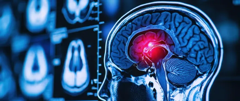
The membranes that surround the brain and spinal cord, known as the meninges, play a crucial role in protecting the central nervous system. However, in rare cases, the cells in these layers may begin to grow uncontrollably, forming a type of tumor known as a meningioma. While meningiomas usually develop slowly and may go unnoticed for years, they can eventually exert pressure on brain tissue, leading to various neurological symptoms.
From vision problems and speech difficulties to memory loss and seizures, meningiomas can manifest across a wide spectrum. If not diagnosed and treated in time, they may result in serious complications. Although most meningiomas are benign, their location and growth rate make careful monitoring essential.
Definition: What Is a Meningioma?
A meningioma is a tumor that originates from the meninges, the protective membranes surrounding the brain and spinal cord. These tumors are typically benign, slow-growing, and often localized, with a higher prevalence in adults, especially women. Meningiomas account for nearly one-third of all brain tumors.
Understanding “what is a meningioma” goes beyond just its medical definition—it involves recognizing its potential effects on surrounding tissues. Although meningiomas rarely invade nearby structures, they can compress adjacent brain regions, leading to significant clinical symptoms. Sometimes, they are discovered incidentally during imaging tests for unrelated conditions. In other cases, they present with signs such as persistent headaches, balance issues, or epileptic seizures.
Origin and Growth Rate of Meningiomas
Meningiomas most commonly begin in the dura mater, the outermost layer of the meninges. While many grow slowly, their effects can vary based on location and size. Some meningiomas remain asymptomatic for years, while others cause increased intracranial pressure and neurological deficits in a shorter period.
Are Meningiomas Cancerous?
Although most meningiomas are non-cancerous, some rare subtypes are classified as malignant. According to the World Health Organization (WHO), meningiomas are graded as follows:
- Grade I: Benign (most common)
- Grade II: Atypical (more aggressive)
- Grade III: Anaplastic/Malignant (highest recurrence risk)
Non-Grade I meningiomas have a higher potential to recur and spread, requiring more intensive treatment and follow-up.
Causes and Risk Factors for Meningioma
The exact causes of meningiomas remain unclear, but certain genetic, environmental, and hormonal factors are believed to contribute.
Primary Risk Factors for Meningioma Development Include:
- Genetic Predispositions: Inherited conditions like Neurofibromatosis Type 2 (NF2) have been linked to a higher risk of meningiomas. Individuals with NF2 may develop multiple meningiomas throughout their lifetime.
- Radiation Exposure: Previous radiation therapy to the head or neck area increases meningioma risk.
- Hormonal Influences: The higher incidence in women and the presence of hormone receptors on some tumors suggest a link between hormones (especially estrogen and progesterone) and tumor growth.
Radiation Exposure and Hormonal Factors in Meningioma Development
The Impact of Radiation
Exposure to high doses of ionizing radiation, particularly to the head, is considered a significant risk factor for developing meningioma. This risk is especially elevated in individuals who were exposed to radiation during childhood, as developing brain tissue is more sensitive to environmental factors.
Hormonal Influences
Meningiomas are more commonly diagnosed in women, suggesting a potential link between female hormones and tumor growth. Estrogen and progesterone receptors have been found in some meningiomas, indicating that these hormones may play a role in stimulating tumor cell proliferation.
Common Signs and Symptoms of Meningioma
Meningioma symptoms can vary widely depending on the tumor’s size, location, and the degree of pressure it exerts on surrounding brain or spinal cord tissue. While some tumors remain asymptomatic for years, others may cause noticeable symptoms even at an early stage.
Symptoms Based on Tumor Location
- Frontal lobe meningiomas may cause cognitive and behavioral changes, such as personality shifts, impaired judgment, and memory problems.
- Tumors located near the cerebellum often lead to loss of balance, coordination issues, and gait disturbances.
- Spinal meningiomas can present with numbness in the hands or feet, muscle weakness, or bladder and bowel dysfunction.
Common Neurological Symptoms of Meningioma
- Headaches are among the most common signs and may intensify in the morning.
- Seizures are frequently observed, especially when the tumor is located near the cerebral cortex.
- Nausea and vomiting may occur due to increased intracranial pressure caused by the tumor's growth.
Visual and Auditory Disturbances
- When a meningioma presses against the optic nerve, it can result in vision loss, double vision, or reduced visual field.
- Tumors located near the auditory nerve may lead to ringing in the ears (tinnitus), hearing loss, or vertigo.
Headaches and Seizures as Warning Signs
Persistent and severe headaches are often a result of the tumor exerting pressure on the surrounding brain tissue. In some cases, patients experience their first-ever seizure, prompting them to seek medical attention—leading to a meningioma diagnosis.

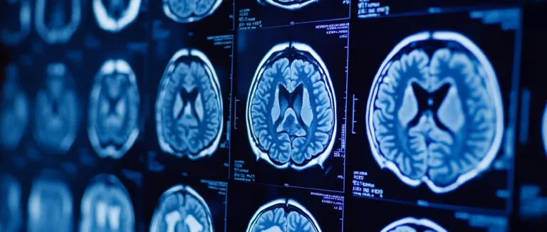
How Is Meningioma Diagnosed?
Meningioma is typically diagnosed through imaging studies performed in response to suspicious neurological symptoms. The diagnostic process involves several key stages: Neurological Examination, Imaging Techniques, and sometimes Biopsy.
Neurological Examination
The diagnostic journey begins with a comprehensive neurological assessment. This includes evaluating the patient’s level of consciousness, reflexes, motor strength, balance, speech, and visual perception. Abnormal findings may raise suspicion of a brain tumor.
Imaging Techniques for Meningioma
- Magnetic Resonance Imaging (MRI) is the most reliable method to assess the size, location, and proximity to surrounding brain structures of a suspected meningioma.
- In some cases, Computed Tomography (CT) scans may also be used, particularly for identifying calcified regions within the tumor.
Biopsy for Definitive Diagnosis
In situations where imaging alone cannot confirm the diagnosis, or when malignant meningioma is suspected, a surgical or needle biopsy may be required. The collected tissue is examined under a microscope to determine the tumor type and grade, which is critical for treatment planning.
Types and Classification of Meningioma
Meningiomas are classified based on their cellular structure, growth rate, and likelihood of recurrence.
World Health Organization (WHO) Classification:
- Grade I (Benign Meningioma): The most common type; slow-growing and typically responds well to treatment.
- Grade II (Atypical Meningioma): Shows abnormal cellular features and has a higher risk of recurrence.
- Grade III (Anaplastic or Malignant Meningioma): Highly aggressive, grows rapidly, and is more likely to recur after treatment.
Common Subtypes of Meningioma
- Fibrous Meningioma: Firm in texture and generally slow-growing.
- Microcystic Meningioma: Contains fluid-filled spaces and has a softer consistency.
- Psammomatous Meningioma: Contains calcified structures, often located near the skull base.
These classifications play a crucial role in formulating an effective meningioma treatment strategy.
Meningioma Treatment Options
The treatment approach is tailored to the tumor’s size, location, and the patient’s age and overall health condition. Available treatment modalities include:
- Surgical Removal
- Stereotactic Radiosurgery
- Radiotherapy
- Chemotherapy (in selected cases)
Surgical Treatment
Surgery is often the first-line treatment. The goal of meningioma surgery is to remove as much of the tumor as safely possible. If the tumor is located near vital brain centers, a partial resection may be necessary. Surgery is particularly effective in Grade I meningiomas and when the tumor's location is favorable.b
Radiotherapy for Meningioma
Radiotherapy may be used when the tumor cannot be fully removed or if it recurs. It is especially employed as an adjunct to surgery in malignant meningioma cases.
Chemotherapy in Meningioma Treatment
Chemotherapy has a limited role in meningioma management. It is typically reserved for Grade III (malignant) tumors or cases resistant to radiotherapy.
Key Factors Influencing Meningioma Treatment Options
WHO tumor grading (Grade I, II, III)
- Tumor location and proximity to critical brain structures
- Risk of recurrence
- Patient’s age, overall health, and comorbidities
What You Should Know About Meningioma Surgery
Meningioma surgery requires meticulous planning and an experienced surgical team. Both preoperative and postoperative phases must be managed with care.
Preoperative Preparation
- Neurological and general health assessments
- Blood tests, advanced imaging, and anesthesia evaluation
- Detailed patient education and consent
Surgical Procedure
The surgery is usually performed under general anesthesia. A small incision is made in the skull to access and remove the tumor. The duration of surgery depends on the tumor’s size and location.
Postoperative Expectations and Recovery
- Hospital stay ranges between 3 to 7 days
- Headaches, fatigue, or mild nausea may occur during the first few days
- Full recovery typically takes weeks to months
- Follow-up MRI scans are used to monitor for recurrence
Robotic Surgery for Meningioma
In select centers, robot-assisted surgery is used for tumors located near the skull base or in challenging regions. This technique allows for greater precision through smaller incisions, improving both safety and recovery time.
Life After Meningioma Treatment
Most patients can return to their normal daily activities following meningioma surgery. However, long-term monitoring and specific care are essential for optimal recovery.
- Neurological follow-ups should be done every 3 months in the first year, then annually
- Routine MRI scans are necessary to detect any recurrence
- Some patients may experience temporary or permanent neurological changes, for which physical therapy and neuropsychological support are recommended
- Long-term neurosurgical follow-up is especially important for Grade II and Grade III meningiomas
Frequently Asked Questions
Yes. Certain types, particularly atypical (Grade II) and malignant (Grade III) meningiomas, have a higher recurrence rate, even after complete surgical removal. Regular monitoring is essential.
A small area may be shaved depending on the surgical approach. However, modern techniques minimize hair removal and prioritize aesthetic outcomes. Hair typically regrows quickly, and any scars tend to fade over time.
Life expectancy depends on tumor type, location, patient age, and overall health. Benign meningiomas usually have excellent outcomes, allowing patients to live many years. Malignant types require more aggressive treatment and close follow-up.
Most meningiomas are benign, but some subtypes can be aggressive. A pathological assessment after diagnosis determines the tumor grade, guiding the treatment plan accordingly.
Small, asymptomatic meningiomas may remain stable or rarely shrink over time. However, active surveillance is crucial, as many tumors eventually show slow growth if left untreated.
* Contents of this page is for informational purposes only. Please consult your doctor for diagnosis and treatment. The content of this page does not include information on medicinal health care at Liv Hospital