Radiology


.webp)


_(1).webp)

.webp)


.webp)
Last Update Date: 9/18/2025 11:16:23 AM
What is Radiology?
Radiology is a medical specialty that examines the structures within the body using imaging techniques and helps to make a diagnosis. Radiologists are medical professionals who specialize in detecting diseases, diagnosing and directing treatment processes using radiological imaging methods. The main purpose of radiology is to visually examine anatomical and pathological features within the body to better understand and diagnose diseases.
What Procedures Does Radiology Perform?
There are different imaging methods that radiologists use. We can list these radiological imaging methods as follows:
- X-Ray: X-rays are used to generate images of bones and some soft tissues within the body. It is widely used for the diagnosis of fractures, lung diseases and many other conditions.
- Computed Tomography (CT): Computed tomography uses X-rays to get detailed 3D images. It is used for detailed imaging of organs, blood vessels and other internal structures.
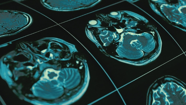
- Magnetic Resonance Imaging (MRI): Magnetic resonance is used to get detailed imagings within the body using strong magnetic fields and radio waves. MRI is widely used especially in the examination of the brain, spinal cord, joints and soft tissues.
- Ultrasonography (USG or Ultrasound): It is used to generate images of organs and tissues within the body using ultrasound waves. It is used in many procedures such as pregnancy follow-up, examination of abdominal internal organs and heart ultrasound.
- Nuclear Medicine: It is used to evaluate the functions of organs and tissues within the body using special substances containing radioactive isotopes. It includes techniques such as SPECT (Single Photon Emission Computed Tomography) and PET (Positron Emission Tomography).
Liv Hospital Radiology
Liv Hospital Radiology Clinic, equipped with high-tech devices, provides healthcare services with a multidisciplinary approach with its specialist physician staff and physicians from different disciplines.
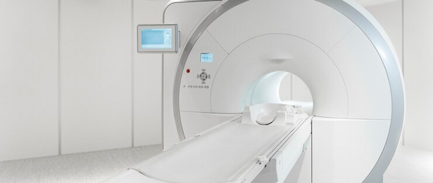
Liv Hospital Radiology Clinic has a 3 Tesla MRI device with a large magnet (70 cm) that is found very rarely in our country, 256-slice CT device, 2 digital x-ray devices, digital fluoroscopy device, 2 portable digital x-ray devices, a digital mammography device, a bone densitometer device, 3 USG and Doppler devices, and an angiography device that guides neuroradiological and vascular interventions.
Low Radiation, Maximum Accuracy at Liv Hospital
Radiological imaging methods are tests that guide physicians in the diagnosis and treatment of diseases. The radiology team provides services to divided regions in the body, for example, some physicians provide services only to the abdomen, some to the brain, and some to the interventional area. All examinations are performed with low radiation doses of up to 80 percent.
MRI Scan in the Comfort of a Room
The internal width of the MRI device in Liv Hospital Radiology Clinic, the ability to broadcast music and videos during imaging are the reasons why the clinic is preferred by patients who are afraid of being trapped in closed spaces or who are overweight. Patients are offered a comfortable, safe environment.
Fully Equipped Approach to Possible Strokes
A patient who had a stroke has a high risk of becoming disabled. At our clinic, the clot that caused the stroke in the patient can be opened with effective interventional radiology techniques without the need for operation. This way, the risk of disability after a stroke is prevented by 100 percent for cases admitted on time. At Liv Hospital Interventional Radiology Clinic, brain aneurysms are treated without operation by entering through the groin. After the procedure, the patient can be discharged immediately.
Bloodless Method for Diagnosing Heart Diseases
In addition to the features found in all CT systems, the 256-slice CT in Liv Hospital Radiology Clinic is a system that can use dual energy technique with its HD detector and single tube and can perform imaging at the most advanced level. Lesion characterization, renal calculus types, detection of hypoperfusion areas in pulmonary embolism examination, metastasis characterization, gout-pseudo gout distinction, reduction of artifacts of metal implants entering the imaging area, plaque characterization of vessels and cardiovascular vessels in the whole body can be performed with advanced imaging techniques.
Calculation of Heart Attack Risk
"Tomographic Coronary Calcium Scoring", another important test performed at Liv Hospital Radiology Clinic, can determine the risk of cardiovascular disease that may occur in the future. Treatment and prevention plans are offered according to the results.
Featured Services
- 3 Tesla MRI
- Computed tomography
- Virtual angiography CT: With virtual angiography, also commonly known as bloodless angiography or 10-second angiography, it is possible to see all the vessels in just seconds, without entering any veins. If no occlusion is detected in the coronary vessels, it provides almost 100 percent diagnostic reliability.
- Vascular interventions
- Digital fluoroscopy
- Digital x-ray
- Bone densitometer
- Digital panoramic cephalometric x-ray
- Color doppler USG
- 4D real time doppler USG